Why is there protein expression after the CRISPR/Cas9 gene editing experiment produces a transcode mutation?
- Hao Shi
- Aug 18, 2025
- 5 min read
CRISPR/Cas9 system is a popular mainstream gene editing technology system with simple operation, high efficiency, and low cost. It can accurately identify and locate specific sequences in the genome or change target sequences in the genome to achieve various purposes, making it a useful tool to study gene function.
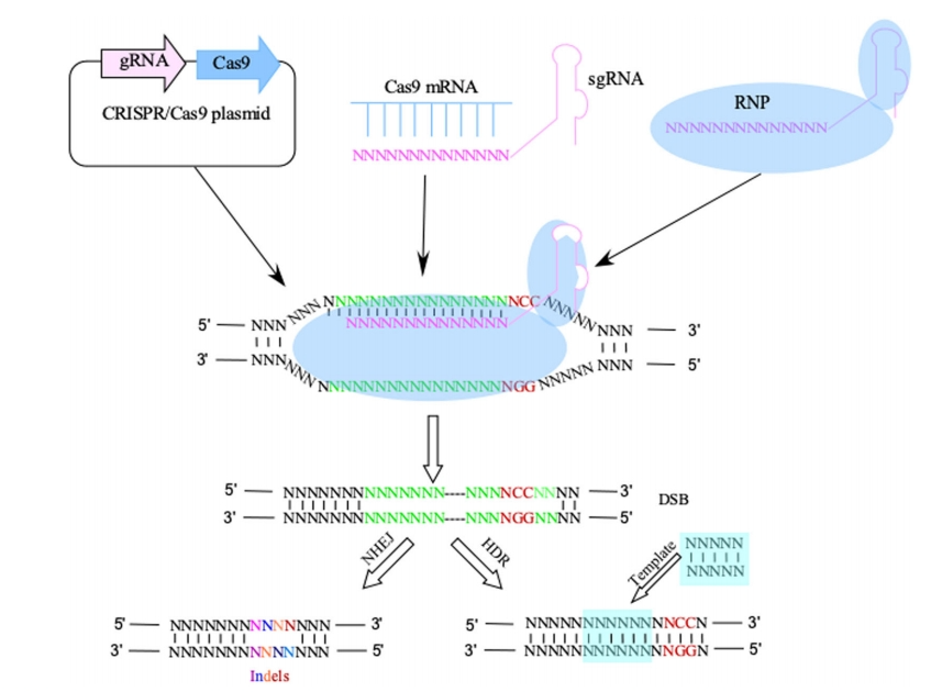
The Cas9 protein, a nuclease enzyme, cleaves DNA single strands to create double-strand breaks (DSBs) at a specific site located by single-guide RNA (sgRNA). Following DNA double-strand breaks, cells typically repair the damage through non-homologous end joining (NHEJ). During this process, mismatches caused by base insertions or deletions may occur, leading to frameshift mutations (Figure 1).

Mutagenesis of codons leads to defects in the mRNA template or translation complex. This triggers quality control (QC) events during translation, which ultimately cause the disintegration of the translation complex. Nonsense-mediated mRNA degradation (NMD), a key pathway affecting CRISPR/Cas9 system gene editing knockout (KO) in mRNA levels, operates through this mechanism.

After the occurrence of a codon shift mutation, it is essential to validate the knockout (KO) results, with DNA and protein level verification being particularly critical. As shown in Figure 3, sequencing data from CRISPR/Cas9 gene editing knockout experiments revealed that the successful KO cell lines exhibited a single base deletion at the sgRNA knockout site compared to the WT/AB wild-type cells, resulting in a codon shift mutation.

(A) sgRNA design sequence;
(B, C) KO successful cell lines and WT/AB wild type Comparison of cell DNA sequence results.
(D) KO successful cell lines and WT/AB Comparison of sequencing results of wild-type cell lines.
DNA-level validation is typically determined by sequencing to detect frameshift mutations, while protein-level validation is usually achieved through Western blotting (WB). When frameshift mutations occur in experiments, we consider gene editing knockout successful. However, it is common to observe residual frameshift proteins despite their disappearance. As shown in Figure 4, compared with the control group, protein bands disappeared in lane 6 while those in lanes 5 remained present, and those in lane 4 were barely detectable. Studies have shown that these residual levels show no significant correlation with mRNA remnants. Although the ideal knockout experiment should demonstrate results like lane 6, why do lane 4 and 5 exhibit such conditions? Let's explore why frameshift mutations at the genetic level may result in complete or partial absence of the encoded protein.

1, 2 and 3 lanes showed the expression level of ITGB1 protein in WT/AB wild-type control group;
4, 5 and 6 lanes showed the expression level of ITGB1 protein in codon shift mutation experimental group
The analysis shows the following reasons:
1. Antibody specificity: Non-specific bands are the most common issue in protein detection. Using high-quality, highly specific antibodies is crucial for obtaining reliable results. If the antibody lacks specificity during experiments, non-specific proteins may appear on the gel strip and interfere with experimental outcomes. Therefore, selecting highly specific antibodies becomes particularly important.
2. Mutation position: A mutation is a DNA chain that has one, two or more bases inserted or missing (not three bases or an integer multiple of three bases), resulting in a corresponding change in the sequence and composition of the codon after the insertion or deletion of the base, resulting in an incorrect amino acid arrangement.
When a codon shift occurs at a position with three or an integer multiple of bases, it may not affect the amino acid sequence. Similarly, when a single base substitution occurs during a codon shift, the codon may alter one of its bases, but the amino acid translated according to the degeneracy of the codon remains unchanged. Both scenarios can result in normal protein expression.
3. Cutting protein: Due to frameshift mutations causing codon shifts, stop codons often appear earlier or later than expected, resulting in elongated or truncated peptide chains. This can lead to residual truncated proteins, which may interfere with antibody binding sites.
4. Experimental operation error: improper operation in the process of single clonal cell sorting leads to 2-3 cells instead of a single cell in one well, and mixed wild-type cells will lead to protein expression or low expression; or the spotting operation error in WB experiment leads to protein expression due to band contamination.
5. Genetic Compensation Effect: The genetic compensation effect refers to the mechanism by which an organism enhances expression of alternative genes to compensate for a gene's complete loss of function after mutation. Two prerequisites for activating this effect are nonsense mutations and nucleotide sequence homology (Ma et al., 2019).
6. Transcript alternative splicing: Alternative splicing refers to the process in which the mRNA precursor transcribed from an exon is produced into different mRNAs by different splicing modes when an exon is destroyed or missing. It is an important mechanism to regulate gene expression and produce protein diversity.
1) Alternative splicing in transcription initiation regions
The site of cleavage during transcription occurs in the transcription initiation region, resulting in the failure of the transcription initiation site to appear at the expected position. When we design sgRNA close to the CDS initiation site, the result is that the sgRNA does not play a role in the expected transcription initiation site and the protein is normally expressed.
2) Exon jumping
The removal of the stop codon may disrupt the splicing rules in the introns. The missing exon sequence is skipped during the mRNA splicing stage and directly connected with the upstream and downstream exons to form a new mRNA, thus continuing the protein translation and leading to normal protein expression (Smits et al., 2019).
Therefore, in gene knockout experiments, the occurrence of a frameshift mutation (current standard for gene knockout) does not guarantee that the target protein will not be expressed or that the gene function will be lost. In this article, we explored some of the reasons why. However, although imperfect, genetic strategies still remain the most powerful means to downregulate target protein expression. Eliminating the protein can serve many useful purposes, including the validation of antibody specificity through gene knockout or gene silencing. At GenuIN, we care to look deeper into these techniques so we can better understand and leverage our proprietary ShGETM gene silencing platform to provide validated shRNA lentiviruses for gene knockdown and validate highly specific antibodies. This allows us to provide reliable resources and services for basic biomedical research and CRO clinical trials.
Reference
MA, Z., ZHU, P., SHI, H., GUO, L., ZHANG, Q., CHEN, Y., CHEN, S., ZHANG, Z., PENG, J. & CHEN, J. 2019. PTC-bearing mRNA elicits a genetic compensation response via Upf3a and COMPASS components. Nature, 568, 259-263.
SMITS, A. H., ZIEBELL, F., JOBERTY, G., ZINN, N., MUELLER, W. F., CLAUDER-MüNSTER, S., EBERHARD, D., FäLTH SAVITSKI, M., GRANDI, P., JAKOB, P., MICHON, A. M., SUN, H., TESSMER, K., BüRCKSTüMMER, T., BANTSCHEFF, M., STEINMETZ, L. M., DREWES, G. & HUBER, W. 2019. Biological plasticity rescues target activity in CRISPR knock outs. Nat Methods, 16, 1087-1093.
TIAN, X., GU, T., PATEL, S., BODE, A. M., LEE, M. H. & DONG, Z. 2019. CRISPR/Cas9 - An evolving biological tool kit for cancer biology and oncology. NPJ Precis Oncol, 3, 8.
ZINSHTEYN, B., SINHA, N. K., ENAM, S. U., KOLESKE, B. & GREEN, R. 2021. Translational repression of NMD targets by GIGYF2 and EIF4E2. PLoS Genet, 17, e1009813.
08/19/2025
Anqi Chen
Supervisor of R&D
GenuIN Biotechnologies


Comments